avm classification radiology
As information has been gleaned a new classification system has emerged that divides vascular anomalies into neoplasms and malformations. MR imaging is the most valuable modality in the classification of vascular malformations.

Medical Radiology Images And Videos Find Free Open Access Medical Content On Grepmed Radiology Radiology Imaging Mri Brain
One postoperative seizure 16.

. 17 They are typically isolated findings although they can be linked with. Spinal AVMs may be classified as intramedullary and extramedullary 80 1 and further divided into four angiographic types with additional subtypes 2-3 see. The Schobinger clinical classification is important to assess patient evolution and indicate intervention.
Its utility is based on accurate initial diagnosis. Vascular tumors are further classified as benign. Intramedullary glomus AVM type III.
Capillary malformations CMs also known as port wine stains have an incidence of 3 per 1000 newborns. Cirsoid AVMs have multiple dilated. It is helpful to compartmentalize AVM angioarchitecture into three components.
The classification of vascular anomalies and their main clinical and MR imaging features are summarized in Table 1. The most recent classification scheme of 2014 continues to divide vascular anomalies into vascular tumors and vascular malformations. At a mean follow-up of 21 months 96 of patients had a modified Engel class I outcome seizure-free 80.
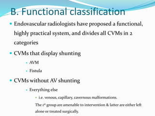
The Schobinger AVM Classification for peripheral AVMs non-neuro is useful to quantify the degrees of symptomatology a patient possesses regardless of the AVMs. Single coiled vessel spinal dural AV fistula type II. It depicts the anatomic relation between the vascular lesion and adjacent.
We recommend to treat symptomatic or evolutive AVMs. 80 1 Or into four types 2. The aim of this review article was to lay out the cor.
Their frequency varies occurring in roughly 10 to 20 persons per 100000. Each of these components have. Spinal arteriovenous malformations can be classified in a number of ways.
Spetzler-Martin classification of cerebral AVM at 4D contrast-enhanced MR angiography and at DSA matched in 18 of 18 patients for both readers which yielded 100. 1 arterial 2 arteriovenous connection and 3 draining veins. We retrospectively reviewed 65.
We believe this generalization to be improper because 1 the classification does not assess the special characteristics of an avm in an individual patient eg associated. Table 1 Clinical and MR Imaging Features of Vascular. 3 This classification has four stages with stage 1.
Hemangioma and arteriovenous malformation AVM are the most commonly used terms and are the mostly incorrectly used as well. 1 Pulmonary AVMs may be classified as simple with a single feeding and draining vessel 80 of cases or complex. Histopathologically AVMs may be classified as cirsoid or cavernous depending on the number and diameter of the intra-lesional vessels.
The Schobinger classification is a clinical assessment of vascular shunting that is predictive of treatment success Table 2.

Spectrum Of Imaging Manifestations Of Vascular Malformations And Tumors Beyond Childhood Radiologic Clinics
Primary Cns Lymphoma Radiology Case Radiopaedia Org Radiology Cns Radiology Imaging

Vascular Malformation Classification References T M O Couto Download Scientific Diagram

Pulmonary Avm Radiology Case Radiopaedia Org Pulmonary Radiology Vascular

Pdf Classification Diagnosis And Interventional Radiologic Management Of Vascular Malformations Semantic Scholar
Frontiers Case Report A Rare Abdominopelvic Arteriovenous Malformation Originating From Splenic Artery And Draining Into Portal Vein

Epos Trade

Pin On Snc

Amar Udare Md Radiogyan Com En Twitter Excellent Talk By Dshatzkes On Vascular Malformation At The Theapdr Noon Conference Take Home Point Be Careful Of What You Call A Hemangioma Refer

Racing Car Sign Radiology Reference Article Radiopaedia Org Radiology Brain Images Medical Imaging

Endovascular Treatment Of Arteriovenous Malformations Of The Head And Neck Focus On The Yakes Classification And Outcomes Journal Of Vascular And Interventional Radiology

Pin On Ct

Stroke Series Video 4 Of 7 Temporal Evolution Of Ischaemic Stroke Presented By Neuroradiologist Dr Frank Gaillard Radiology Mri Mri Brain

References In Pulmonary Arteriovenous Malformations And Their Mimics Clinical Radiology
Cerebral Pial Arteriovenous Malformation Avm
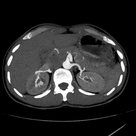
Renal Arteriovenous Malformation Radiology Reference Article Radiopaedia Org

The Borden Classification Of Dural Arteriovenous Fistulas Davf Groups These Lesions Into Three Types Based Upon The Site Of Ve Borden Classification Proposal

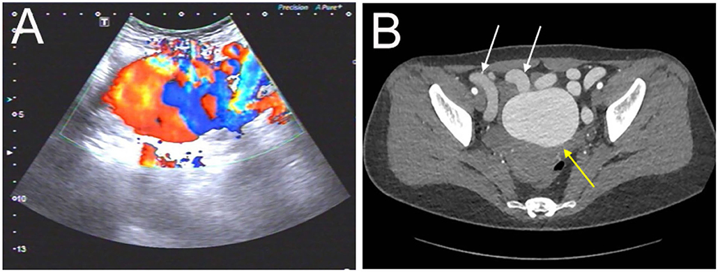
Uterine Arteriovenous Malformation Radiology Case Radiopaedia Org

Diagnostics Free Full Text Imaging Features Of Post Main Hepatectomy Complications The Radiologist Challenging Html


Comments
Post a Comment